Your eyes maintain a delicate balance of fluid pressure that keeps them healthy and functioning properly. When this balance is disrupted, it can lead to glaucoma, a condition that gradually damages the optic nerve.
The challenge is that you won’t feel high pressure or notice early nerve damage on your own. Detecting glaucoma requires specialized equipment and expertise that only your eye doctor can provide during a comprehensive exam.
Keep reading to learn about the different tests that work together to identify glaucoma and protect your vision.
How Is Glaucoma Diagnosed?
Diagnosing glaucoma requires more than just one test. Your eye doctor combines several different evaluations to get a complete picture of your optic nerve health and visual function. Each test provides specific information that helps confirm whether glaucoma is present and how advanced it may be.
Optic Nerve Examination
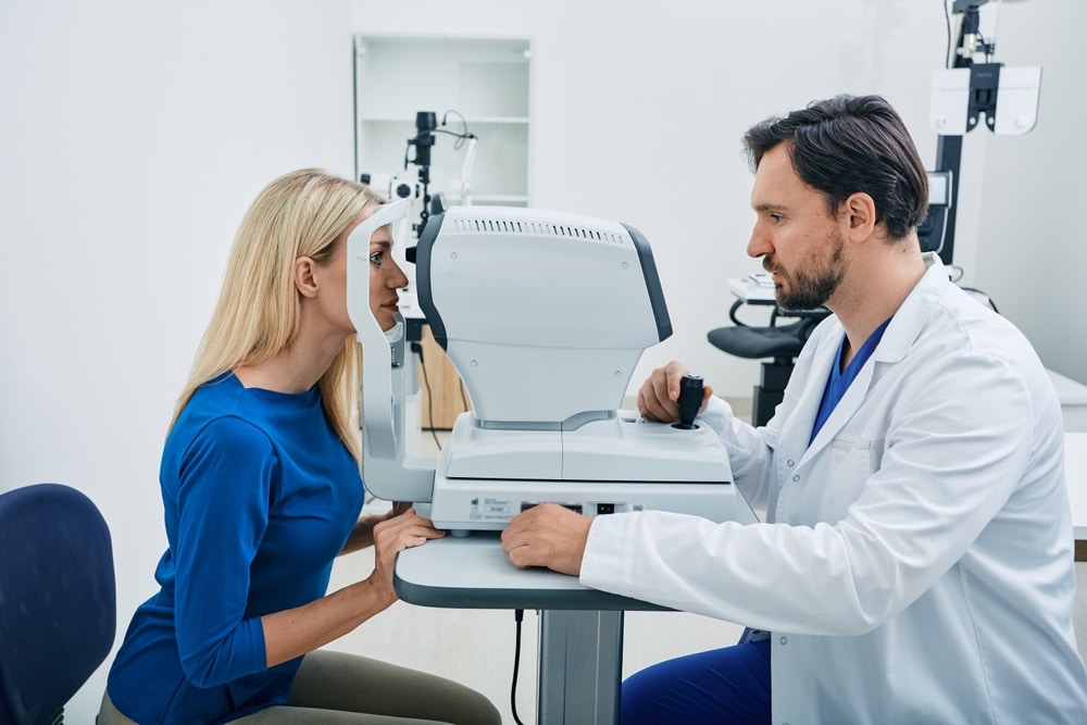
Your eye doctor examines the optic nerve at the back of your eye using a special magnifying lens. This part of the exam typically requires dilating drops to widen your pupils for a better view.
They will look for specific signs of damage, including changes in the nerve’s appearance and a condition called “cupping,” where the center of the nerve becomes hollowed out. These visual changes indicate that glaucoma may be damaging the nerve fibers that transmit information from your eye to your brain.
Eye Pressure Testing
Tonometry measures the fluid pressure inside your eye. During this test, your doctor may use different methods depending on the equipment available.
There are different ways to test eye pressure, such as the quick air puff test and applanation tonometry, which uses a small probe that gently touches the surface of your eye after numbing drops are applied.
Normal eye pressure typically ranges between 10 and 22 millimeters of mercury, though some people develop glaucoma even with normal pressure readings.
Visual Field Examination
This test checks your peripheral vision, which is often the first area affected by glaucoma. You’ll look into a machine and press a button whenever you see small lights appear in different locations.
The test maps out your complete field of vision and identifies any blind spots that might indicate nerve damage. These assessments typically take about 10 minutes per eye and provide detailed information about how well you can see in all directions.
Additional Testing (OCT)
Optical coherence tomography, or OCT, uses light waves to create detailed images of your optic nerve and the surrounding tissue. This advanced technology measures the thickness of your nerve fiber layer with incredible precision. OCT scans help doctors detect even subtle changes in nerve health and monitor whether glaucoma is progressing over time.
How Often Should I Have a Glaucoma Screening?
The frequency of glaucoma testing depends on your age and risk factors.
Most adults should have a baseline screening by age 40, with follow-up exams every two to four years if no risk factors are present. After age 60, annual screenings become more important as glaucoma risk increases.
However, you may need more frequent testing if you have a family history of glaucoma, are of African or Hispanic descent, have diabetes, or take steroid medications.
Your eye doctor will recommend a personalized screening schedule based on your individual risk profile.
Protect Your Vision With Regular Eye Exams
Early detection makes all the difference when it comes to glaucoma. These comprehensive tests work together to identify problems before you notice any vision changes.
Schedule a comprehensive eye exam at All Eye Care PA in Waxahachie, TX, today to protect your vision with thorough glaucoma screening and personalized care from experienced eye doctors.